The purpose of this illustration is to teach medical students the spatial relationships between the nerves, arteries, and muscles within the infratemporal fossa. These structures are situated closely together in this cavity and are usually illustrated separately.Three views were chosen to capture the layered complexity of this cavity. The main superficial view shows the relationship between the maxillary artery and the lateral pterygoid muscle. It also depicts the relationship between the maxillary artery’s branches and the nerves arising from the mandibular branch of the trigeminal nerve. In the deeper view, the lateral pterygoid muscle is removed to reveal the mandibular branch exiting through the foramen ovale. Particular attention was paid to how the nerves and arteries interweave between the pterygoid, buccinator, and temporalis muscles to reach their target structures through research and cadaveric dissection. The coronal section illustrates how the two heads of the lateral pterygoid muscle lay within the concavity of the fossa, and how the lateral and medial pterygoid muscles are positioned relative to the lateral pterygoid plate.
CT segmented with Horos for anatomical plate reference.
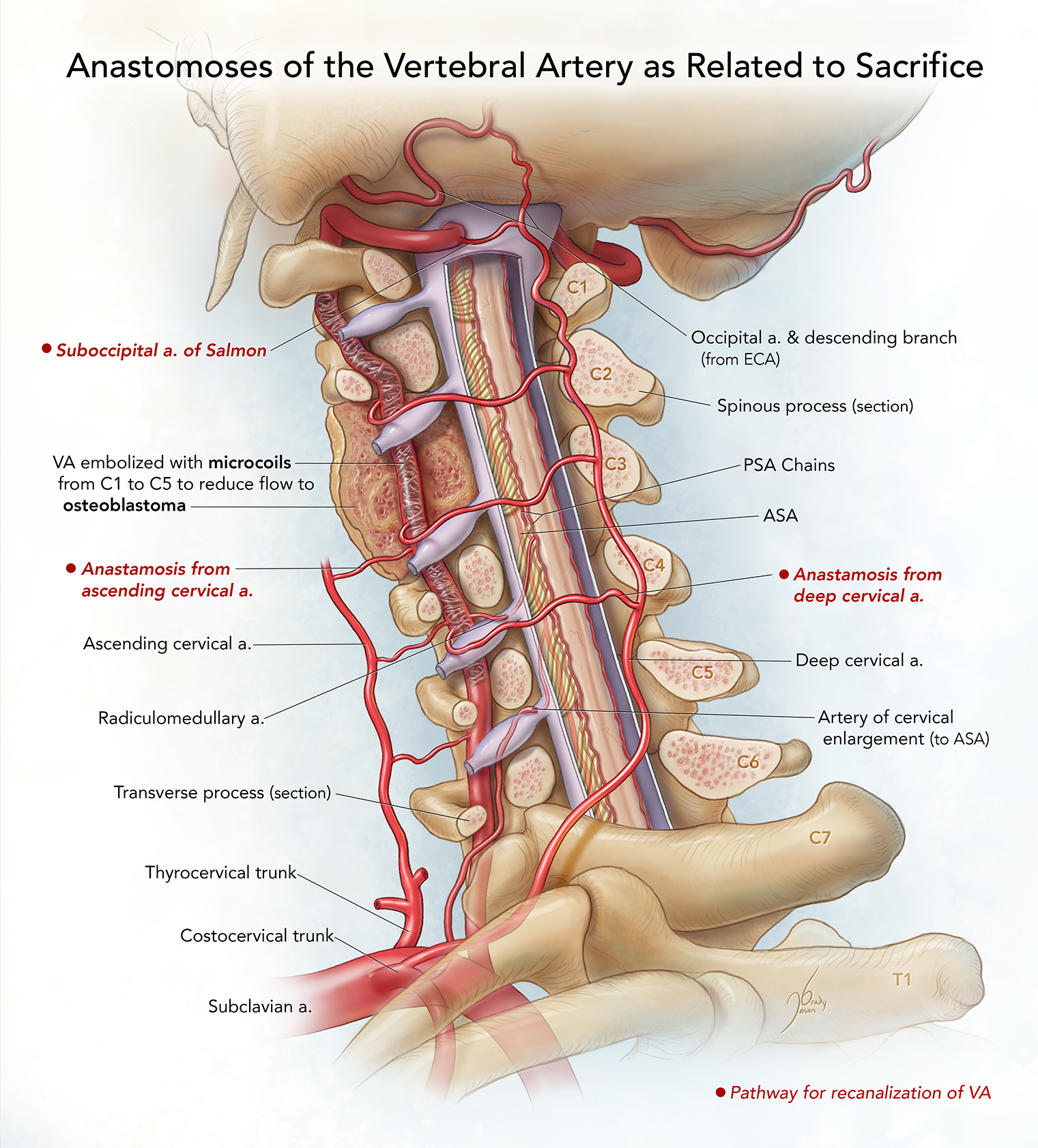
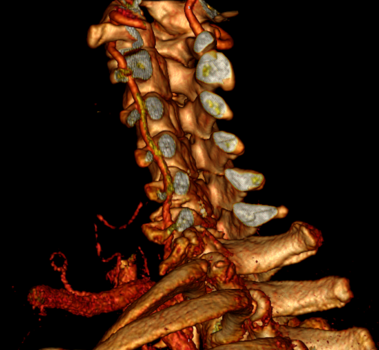
Vertebral Artery Sacrifice
This illustration depicts anastomoses of the vertebral artery (VA) that are relevant to VA sacrifice, which involves embolizing the VA with metallic microcoils across multiple cervical levels. In this case, the VA was sacrificed to reduce blood flow to an osteoblastoma. The anastomoses (red labels) from the suboccipital artery of Salmon, ascending cervical, and deep cervical arteries may recanalize the VA after sacrifice. The transverse and spinous processes, vertebral pedicles, and dura are sectioned to reveal the spatial relationships of the arterial supply (bold italic labels) of the spinal cord to the VA, which must be considered during VA sacrifice to avoid spinal cord damage.
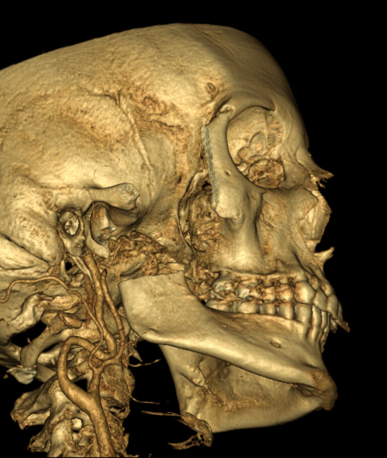
CTA segmented with Horos for anatomy surrounding vertebral artery.
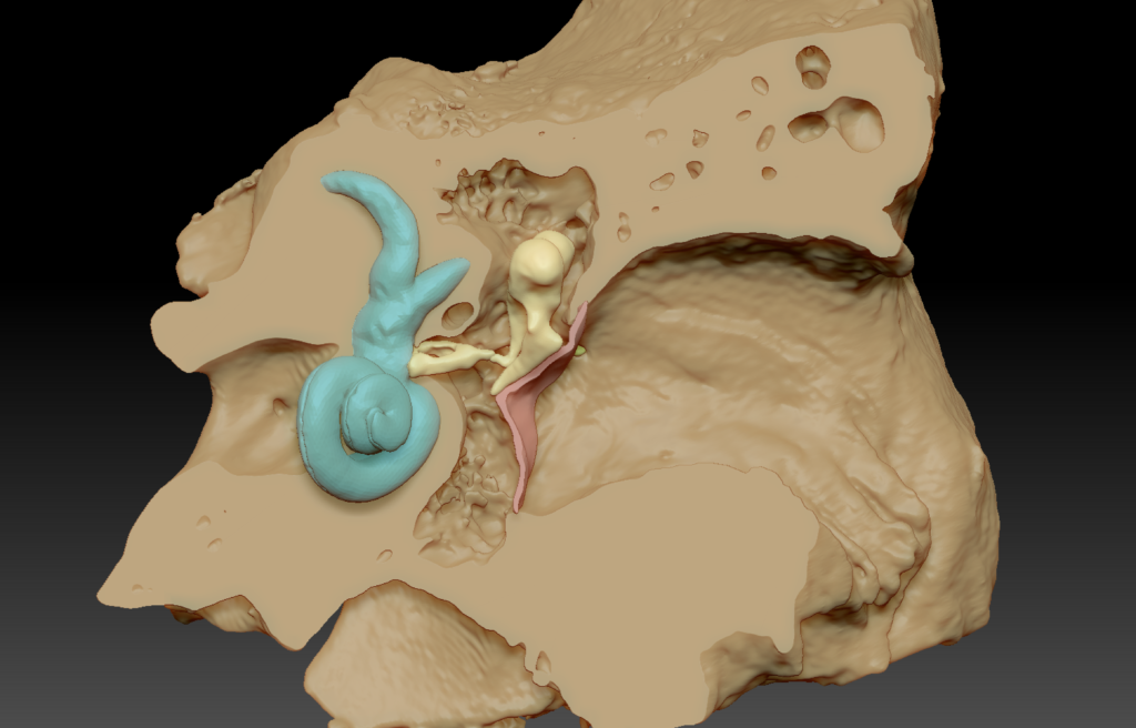
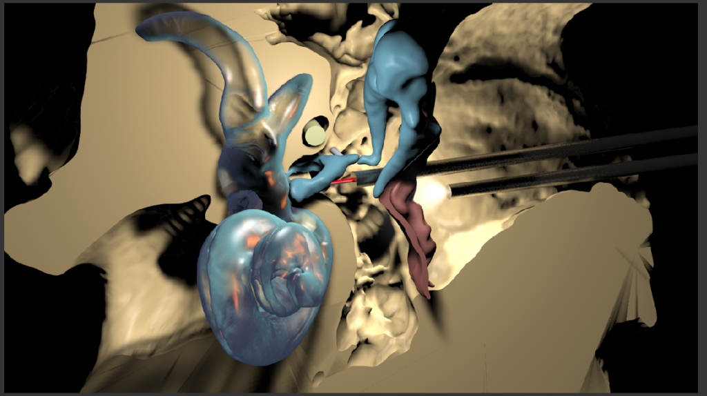
Stapedectomy
This illustration depicts a stapedectomy. A microCT segmentation of the temporal bone was used as reference. A boolean was used in ZBrush to achieve a cross section to show the spatial relationships of important structures. This illustration depicts an endoscopic approach in which the anterior crus of the stapes is divided with a laser.
ZBrush Boolean of MicoCT of temporal bone as well as the Cinema4D scene with instruments for Stapedectomy.